
Which Ultrasound Machine Is Best for Mobile Clinics?





A mobile clinic often works where space, power access, staffing, and patient flow are limited. A unit may move between community sites, employer health programs, rural outreach locations, temporary screening events, or home-visit services. In that setting, an ultrasound system that performs well in a fixed imaging department can become impractical if it is too heavy, slow to start, difficult to clean, or dependent on mains power.
Which ultrasound machine is best for mobile clinics? For most mobile services, the best choice is not simply the smallest device. It is a portable or compact cart-based system that matches the clinic’s core examinations, provides dependable image quality in its required depth range, supports the necessary probes, and can operate through a realistic workday with stable battery and connectivity options. Handheld ultrasound may be the best fit for focused point-of-care examinations, while a premium portable laptop-style system is usually more suitable when the clinic provides broader abdominal, obstetric, vascular, or musculoskeletal services.
Start with the examinations the clinic must actually perform
The most costly purchasing mistake is selecting a machine based on portability before defining the clinical workload. “Mobile” does not describe one uniform use case. A team performing focused cardiac checks at bedside has different needs from a travelling service providing scheduled pregnancy scans, abdominal imaging, or lower-limb vascular assessments.
Begin by listing the examinations that will make up most appointments, then identify which findings must be visualized reliably. This clarifies whether the system needs high-frequency superficial imaging, deeper penetration, color and spectral Doppler, cardiac presets, needle guidance tools, measurement packages, or formal reporting functions.
- Focused assessments: Handheld or tablet-connected ultrasound may be appropriate for rapid lung, bladder, basic abdominal, vascular access, soft-tissue, and limited cardiac assessment when the clinical protocol supports focused imaging.
- General primary-care imaging: A portable laptop-format machine with convex and linear probes is often a practical starting point for abdominal, pelvic, thyroid, vascular access, and musculoskeletal work.
- Obstetric services: Image quality, obstetric calculations, fetal measurements, appropriate probe selection, and a screen large enough for reliable assessment generally matter more than minimum device weight.
- Vascular work: Color Doppler, pulsed-wave Doppler, image optimization controls, a high-quality linear transducer, and measurement workflow are central. A basic handheld device may not meet the service requirement.
- Cardiac examinations: A phased-array probe, cardiac software, adequate frame rate, Doppler capability where needed, and clinician familiarity with the system should be confirmed before purchase.
A mobile clinic does not need every available application package. It does need a device that can perform its highest-risk or most frequent examination consistently. Buying a compact system with insufficient imaging depth or missing Doppler functions can force referral of the very cases the service was intended to assess.
Choose the format before comparing specifications
Portable ultrasound systems generally fall into three practical categories. Each can be useful, but their operating limits differ.
Handheld systems are appealing because they fit easily into a clinical bag. That convenience is real, especially where examinations are short and targeted. But a small device can transfer practical burdens to the operator: managing a separate phone or tablet, maintaining device security, checking wireless connections, protecting the screen from glare, and ensuring that the application workflow meets local documentation requirements.
A laptop-style portable system is often the more balanced option for a mobile clinic with scheduled appointments. It can usually support more probes and imaging modes without requiring a full-sized cart. Its larger screen also matters when clinicians need to evaluate subtle boundaries, make measurements, or review images with a patient.
Compact cart systems suit clinics that travel by vehicle but perform a high number of examinations at each stop. Their mobility depends less on device weight and more on whether the clinic can safely unload, roll, position, clean, recharge, and secure the machine at every location.
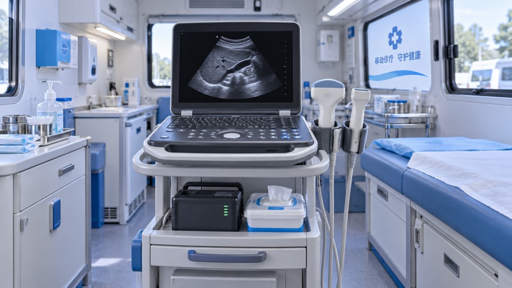
Image quality should be tested under mobile-use conditions
Brochure images are not enough. A system may produce attractive images under ideal settings but be less effective when the operator is working in a bright temporary room, assessing a larger patient, scanning through difficult acoustic windows, or moving quickly between appointments.
During evaluation, ask for demonstrations using the examinations most relevant to the service. The review should cover shallow structures as well as deeper targets where applicable. Assess border definition, penetration, noise, color sensitivity, frame rate, and the responsiveness of controls. If Doppler is clinically necessary, do not treat it as an optional feature merely because color flow appears on the screen; confirm the relevant Doppler modes, measurement tools, and practical image quality.
Screen visibility deserves separate attention. Mobile sites may have uncontrolled lighting, reflected daylight, or limited ability to dim a room. A bright, adequately sized display reduces the temptation to scan in poor viewing conditions. Touchscreen-only controls can be convenient, yet physical controls may be faster when operators need to adjust gain, depth, focus, or Doppler settings while maintaining probe position.
Battery claims matter less than the complete operating cycle
Battery life is frequently judged by a single number, but real endurance depends on screen brightness, Doppler use, probe activity, wireless transmission, standby periods, and repeated powering on and off. A clinic should consider the full cycle: travel, setup, patient scanning, image review, documentation, cleaning, packing, and transport to the next site.
Ask practical questions rather than relying only on nominal run time:
- Can the battery be changed without interrupting workflow?
- How many batteries are needed for a normal clinic session?
- Does the system charge in a vehicle, from AC power, or through a docking arrangement?
- How long does recharge take after a full day of use?
- What happens if power is lost while images are being saved or transferred?
- Are chargers, spare batteries, and cables easily available through the intended supply channel?
A mobile clinic should avoid building its schedule around a battery estimate with no margin. Where power is uncertain, a spare battery or a clear charging routine may be more valuable than selecting a slightly lighter system. The team also needs a storage and transport process that prevents batteries from being exposed to unsuitable heat, impact, or moisture.
Probe strategy is more important than probe quantity
Every additional transducer expands clinical capability, but it also adds cost, cleaning work, packing complexity, and the possibility that an essential item is left behind. The appropriate probe set follows the examination list, not a desire to cover every specialty.
A convex probe is commonly needed for abdominal and many obstetric applications. A linear probe is useful for vascular access, superficial structures, thyroid, soft tissue, and many musculoskeletal examinations. A phased-array probe may be needed for cardiac work. Endocavitary imaging introduces additional requirements for compatible covers, cleaning and high-level disinfection processes where applicable, privacy arrangements, and safe probe handling.
Probe durability should be reviewed alongside image performance. In a fixed department, probes may remain attached to the system. In a mobile clinic, they are repeatedly connected, disconnected, packed, unpacked, and moved through doorways and vehicles. Check connector protection, cable strain relief, carrying arrangements, and whether the system has a secure place for each probe during transport.
Workflow problems often appear after the scan, not during it
Obtaining an image is only one part of mobile ultrasound work. The clinic must identify the patient correctly, store the examination securely, document findings, transfer images where required, and maintain continuity when the network is unavailable. A highly portable device can create risk if these steps rely on informal workarounds.
Before deciding, map the expected workflow from registration to record completion. Confirm how patient identifiers are entered, whether images can be reviewed later, which formats can be exported, and how the system handles temporary loss of internet access. Some services need integration with an existing clinical record environment; others need a secure standalone archive and a reliable process for later upload. The choice should fit the actual operating model rather than assume constant Wi-Fi or mobile data coverage.
Cybersecurity and access control also belong in the evaluation. Shared mobile devices should have role-appropriate access, secure sign-in procedures, and a defined approach for lost or stolen equipment. Software update practices matter because a device used in the field can be missed if updates require a return to a central site.
Transport, cleaning, and ergonomics can determine whether the system remains usable
A machine may be technically portable but still unsuitable for repeated outreach work. Consider the route from vehicle to examination area: stairs, narrow corridors, gravel, wet entrances, lifts, and temporary rooms with limited space. Evaluate cases, wheels, handles, cable storage, and the time required to set up safely.
Cleaning procedures should be realistic between patients. Smooth, wipeable surfaces and accessible probe holders reduce delays. The clinic must use cleaning and disinfection methods compatible with the manufacturer’s instructions for the system and transducers. Improvised cleaning products or poorly controlled probe storage can shorten equipment life and create infection-control concerns.
Ergonomics affect both image quality and staff fatigue. A system that forces the operator to bend over a low table, twist toward a side-mounted display, or hold a tablet at an awkward angle can make consistent scanning difficult. Test the device in a setup similar to the intended clinic environment, including typical chairs, examination couches, and lighting.
A practical selection path for procurement teams
Rather than comparing a long list of features, create a short decision matrix based on the clinical service. Give higher priority to functions that cannot be replaced by operator technique or accessories. For example, a vascular service should place Doppler performance and linear probe quality ahead of minimum device weight. A home-visit team delivering focused assessments may rank startup speed, battery handling, and carrying convenience more highly.
- Define the core examination list and the findings that must be visible.
- Set minimum requirements for probes, imaging modes, measurements, reporting, and data storage.
- Decide whether the service needs a handheld, laptop-style, or compact cart platform.
- Test candidate systems in realistic lighting, power, connectivity, and space conditions.
- Review the full accessory package, including batteries, chargers, cases, probe holders, and cleaning compatibility.
- Confirm service support, warranty scope, repair process, software maintenance, and availability of replacement probes in the intended market.
- Train users on both scanning workflow and the less visible tasks of patient identification, image transfer, cleaning, packing, and fault reporting.
The best ultrasound machine for a mobile clinic is therefore the one that supports the clinic’s actual level of diagnostic responsibility without creating avoidable gaps in power, documentation, transport, or infection-control workflow. A compact handheld device can be an excellent focused-assessment tool. A portable laptop system is often the stronger choice for mixed diagnostic work. Where appointments are more complex or volume is high, a compact cart-based platform may justify the additional transport effort because it provides a more stable working environment for the operator and patient.
Related News
- Which Ultrasound Machine Is Best for Mobile Clinics?Oct 13, 2026Which ultrasound machine is best for mobile clinics? Compare handheld, portable, and cart-based systems for imaging quality, battery life, probes, workflow, and reliable care.
- U.S. Requires New NMPA Classification Certification for Exported Medical Ultrasound SystemsAug 16, 2026U.S. requires new NMPA classification certification for exported medical ultrasound systems. Learn the latest compliance changes, export risks, and what companies must check now.
- China Tightens Medical Device Export Compliance for Ultrasound SystemsAug 15, 2026China tightens medical ultrasound export compliance from Sept. 1, 2026. Learn the new NMPA filing rules, risks, and what exporters must do now.
- EU CE Rule Takes Effect for Ultrasound EMC TestingAug 08, 2026EU CE Rule Takes Effect for Ultrasound EMC Testing: learn how EN IEC 60601-2-37:2026 reshapes CE compliance, EMC reports, AI module testing, and EU market access for ultrasound systems.
Related News
- 00
0000-00
Medical Imaging Trends Shaping Diagnostic Investments in 2026 - 00
0000-00
Dental Laser Systems: When the Upgrade Delivers Real Clinical Value - 00
0000-00
Smart Hospitals Technology Trends Shaping Care Delivery in 2026 - 00
0000-00
Precision Medical Imaging in 2026: Performance Trends That Matter - 00
0000-00
Clinical Integration Solutions for Hospitals: Reducing Data Gaps and Workflow Risk
Author :
Industry Global
Weekly Insights
Stay ahead with our curated technology reports delivered every Monday.